A sensor unit for non-invasive detection and analysis of the pulsating blood flow waveforms by means of the reflective single-period photoplethysmography (SPPPG) technique has been designed and clinically tested. The sensor is operated jointly with any standard PC, by connecting the sensor head to the AD-card and using a separate hard disc with the signal processing software; all circuits are feeded by the PC power supply. After processing, normalized shape of the mean SPPPG signal and its parameters are calculated and displayed; the measurement/processing time does not exceed 2 minutes. The clinically detected SPPPG signal shapes and corresponding parameters are presented and discussed. The preliminary results confirm good potential of this sensing approach for fast and patient-friendly early cardiovascular diagnostics.
1. INTRODUCTION
Photoplethysmography (PPG) is a non-invasive method for studies of the blood volume pulsations by detection and temporal analysis of the tissue-scattered absorbed optical radiation. Blood pumping and transport at different body locations - fingertip, earlobe, forehead, forearm, etc. - are monitored with simple and convenient PPG contact sensors.
When the tissue is illuminated by visible or near-infrared radiation, heartbeat-period changes in the transmitted and scattered optical signal levels can be recorded by means of the photoplethysmographic (PPG) sensors [1] (see Fig. 1). The PPG signals are originated by absorption of optical radiation by the pulsating blood volume, therefore they contain clinically valuable information on the blood pumping and transport conditions in living body.
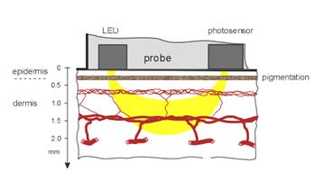
Fig. 1. Placement of optical Devices
A number of transmission-type finger and earlobe PPG devices for monitoring of heartbeat rate and tissue blood supply have been designed and routinely used. Advanced designs of the back-scattering or reflection-type PPG sensors [2] are of increased interest today, mainly thanks to the their clinically more convenient one-touch operation mode. However, the reflected PPG signals are weaker and therefore more noisy, than the transmitted ones.
Full and clear clinical interpretation of all components of the PPG signals is still problematic. Qualitatively, one can assume that the initial part of the detected heartbeat signal (raising front and systolic peak) mainly reflects the heart condition and activity, while the following part of a pulse is generally determined by elasticity and other features of the vascular system (see Fig. 2).
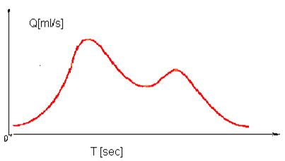
Fig. 2. PPG signal
PPG signals are not strictly repeating and periodical, there are slight fluctuations of the signal amplitude, baseline and period [3]. Consequently, the fluctuations take place relatively to some virtually stable mean single-period PPG (SPPPG) signal. This mean signal can be identified by averaging a number of sequent PPG pulses over a time interval longer than 50 seconds (which is the longest fluctuation period [3]). In our project developed technique of the reflected PPG signal accumulation and integrated processing made possible to detect and analyze the mean SPPPG signals with fairly good accuracy and quality [4, 5]. Following initial results, the mean SPPPG signal shape appears to be a very individual feature for each monitored person; qualitative differences in signal shapes for healthy individuals compared to those for persons with cardio-vascular disorders were observed. Obviously, the SPPPG signal shape contains certain coded information regarding the cardio-vascular state of the patient, and a detailed shape analysis eventually might provide clinical data for early cardio-vascular diagnostics in future. To check this opportunity in clinical environment for larger number of patients, more specific sensor design and signal processing technique had to be developed. This paper describes the design and signal processing concepts of developed PPG sensor as well as some results of clinical trials carried out with this device.
2. DESIGN OF THE UNIT
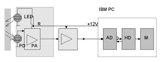
The basic design of the unit is relatively simple. It consists of optical contact probe, bio-signal amplifying/filtering circuit (both powered by a rechargeable battery) and a lap-top computer with specially developed software for AD-conversion, storage, processing and display of the PPG signals. All equipment is placed in a hand-held case .The advanced SPPPG sensor is intended for clinical use in conjunction with any standard PC. Three basic modules are needed for its operation - the sensor head (fingertip probe with amplifier), standard AD-card and a standard hard disk (preferably separate) with the signal processing software and space for storage of the recorded data. Feeding of all electronic circuits is provided by the PS power supply (see Fig.3).
Tissue Contact probe Amplifier AD-card Hard disc Monitor
Fig. 3. Block-diagram of the optical PPG sensor unit
Block-diagram of the device is presented on Fig. 3. The finger contact probe - sensor head - comprises a continuously emitting diode LED (GaAs, lmax ~ 940 nm) integrated with a photodiode PD (Si, 1 cm2 active area) see Fig.3 and a pre-amplifier chip PA. The pre-amplified PD output signal was passed via a flexible cable to the broadband amplifier and further to the AD-card input. Only the ac component of the photodiode output signal was amplified and further processed; the amplifier provided about 400-fold magnification. Following results of recent study [4], frequency filtering of the remitted PPG signals may cause their shape-deformations, therefore signals of all frequencies were amplified and passed to input of the 12-byte AD-card.
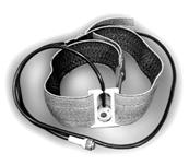
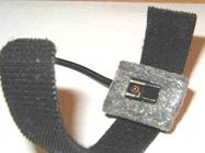
Fig. 4. The PPG contact probe
The LED and photodiode of the fingertip probe were down-oriented during the measurements, and additional calibrated load was applied to provide equal pressure force (0.65 N) to the fingertip skin for all monitored patients. The contact probe was placed within a vertical cylindrical capsule, which served simultaneously as a finger-holder, external light shield and sliding guide for the optical probe. The 3rd (middle) finger of left arm was mainly used for the PPG signal recording, and the patients were kept in horizontal position (lying on their back) during the measurements.
3.THE SIGNAL PROCESSING
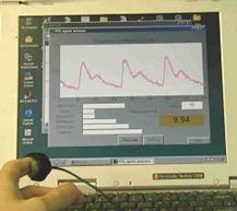
The signal processing software was stored on a separate hard disk, which served for operation of the sensor device and for storage of the measured and calculated data. The algorithm for integration and averaging of the detected PPG signals [4] was updated following our recent experience, and a service program was added. At the beginning of each trial, a window for entering the monitored patient data (name, age, pathology, etc.) was opened. Then a measurement window with instructions appeared, and after proper placing of the fingertip probe the measurements were started. The data were recorded for 60 - 80 seconds, then the whole PPG signal was stored in the HD memory and processed.
A special algorithm calculated the mean normalized SPPPG signal for the monitored person and the corresponding signal shape parameters (maxims, minims, amplitude ratios, integral area, etc.). Heartbeat rate and arrhythmia could be calculated, as well. All these data appeared on the PC monitor within less than 5 seconds, so the time necessary for the whole procedure normally did not exceed two minutes.
4.THE SOFTWARE PROGRAMING
Special software was developed for the PPG bio-signal acquisition, processing and data storage, offering the following options [5-6]:
- Filling the first window for patient data - name, age, gender, complains, doctor´s comments, etc.;
- Pre-setting of the measurement time schedule;
- The PPG signal registration and display in real time;
- Signal clean-up (special filtering algorithm) and calculation of the mean single-period PPG (SPPPG) signal shape;
- Calculation of specific cardio-vascular parameters for the registered signals - heartbeat rate, anacrota rise-time, time delay and relative amplitude of the secondary peak (dycrotic notch), etc.;
- Display of the corresponding PPG parameter set with subsequent cardio-vascular assessment results;
- Storage of the measurement/assessment data.
5. THE PPG CONTACT PROBE
The optoelectronic contact probe continuously emits radiation into the under-skin tissues with blood vessels and detects the AC-component of the back-scattered radiation that reflects the blood volume pulsations.
a b
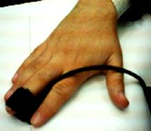
Fig.4. The PPG contact probe (a) and its application for the fingertip monitoring (b)
The probe comprises a GaAs emitting diode (diameter of the emitting area ~2 mm, power ~10 mW, peak wavelength ~940 nm), and a Si photodiode with square detection area ~5x5 mm). Both diodes are closely mounted on a soft plastic pillow and fixed onto the measurement site by means of a sticky band - see Fig. 4,b. The band length is adjusted to the fingertip measurements (Fig. 4, a); however, the band easily can be extended by spare bands, if necessary, so providing possibility to take PPG measurements from different locations of the body, forehead, neck, forearm, knee (Fig. 5).
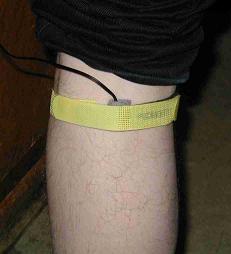


Fig. 5. Application of the PPG contact probe at forehead, neck, forearm and knee
6. THE MEASUREMENT RESULTS:
SOME EXAMPLES
The developed bio-sensor unit had undergone several tests, and some interesting clinical results were obtained; they will be presented and discussed below. Most of these measurements were taken from the middle fingertip of the left hand.
We observed and recorded several abnormalities of heart function, including partial or total lack of one heartbeat in the cardiac sequence (see fig. 6). Typically, the next heartbeat after the missing one is more intensive than others in the sequence, so obviously the heart is auto-compensating the short-term lack of blood pumping. The monitored persons did not feel any discomfort during the missing heartbeat. This phenomenon was recorded several times, so there was little doubt that both persons had trouble with heart functioning, and they were recommended to visit cardiologist for further investigations.
Consequently, the PPG sensor unit appeared helpful for early warning of cardio-vascular dysfunction, so it seems to have good potential for primary cardio-vascular assessment and early screening of the risk patient groups in future.


a b
Fig. 6. The observed heartbeat irregularities for two monitored persons
7. SUMMARY
- A small-size portable PPG sensor device (44×32×9 cm, 4,1 kg, battery-powered) is designed, constructed and tested.
- The contact probe design provides reliable pulsating blood flow measurements at different sites of the body.
- Several interesting clinical conditions have been recorded with the device by temporal analysis of the PPG signals - heartbeat irregularities, arrhythmic and spasmatic responses to intensive physical exercises, right-left hand fingertip blood flow differences, etc.
- Shapes of mean SPPPG signals recorded at various locations of the body contain valuable cardiovascular information.
- The proposed approach and sensor design proved to be suitable for fast primary cardiovascular assessment and early screening.
REFERENCES
- A. B. Hertzman. "Photoelectric plethysmograph of the finger and toes in man", Proc. Soc. Exp. Biol. Med., 37, pp. 1633-1637, 1937.
- H. Ugnell. "Phototplethysmographic Heart and Respiratory Rate Monitoring", Ph. D. Thesis No. 386, Linkoping University, 2002.
- M. Nitzan, H. de Boer, S. Turivnenko et al. "Power spectrum analysis of spontaneous fluctuations in the photoplethysmographic signal", J. Bas. Clin. Physiol. Pharmacol., 5, No. 3-4, pp. 269-276, 1994.
- J. Spigulis, U. Rubins. "Photoplethysmographic sensor with smoothed output signals", Proc. SPIE. 3570, 2003, pp. 195-199.
- M. Ozols, J. Spigulis. "Acquisition of biosignals using the PC sound card", Proc. Int. Conf. "Biomedical Engineering" (KTU, Kaunas, LT), pp. 24-27, 2001.
- M. Ozols, J. Spigulis. "Analog-to-digital conversion of bio-signals by means of the PC sound card", Proc. Baltic Electronics Conference BEC´2002 (TTU, Tallinn, EE), 2002.


